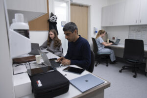
The Women’s Health Research Institute (WHRI), on behalf of the Skidmore Goodman Perinatal Research IMaging and Evaluation (PRIME) Centre, and in partnership with the BC Women’s Health Foundation, is pleased to congratulate the recipient of the 2026 PRIME Centre Research Award.
The Skidmore Goodman PRIME Centre is an ultrasound imaging research and teaching facility within the Skidmore Goodman Research Centre for Women’s Health at BC Women’s Hospital + Health Centre. The PRIME Centre is dedicated to the development of new imaging research and technology to benefit women’s, fetal, and neonatal health. The centre provides researchers with access to imaging equipment and offers opportunities for training in ultrasound technical skills.
One award, valued at $30,000, was distributed for a one-year period. The Skidmore Goodman PRIME Centre Award provides funding for research in women’s, fetal and/or neonatal health research. The research must be conducted in the Centre and makes use of the available equipment, facility features and personnel support available, while emphasizing collaborative projects between university departments, medical specialties, and industry in accordance with CFI guidelines.
Recipients

PROJECT TITLE:
Can Quantitative Ultrasound detect Adenomyosis in individuals with Abnormal Menstrual Bleeding?
PRINCIPAL INVESTIGATOR:
Dr. Mohamed Bedaiwy, Professor, Department of Obstetrics and Gynecology, Faculty of Medicine, University of British Columbia
Robert Rohling, Mady Salker, Farah Deeba, Shahed Khan Mohammed
SUMMARY:
Adenomyosis is a common but often underdiagnosed condition where the womb lining grows into the womb’s muscular wall. This leads to painful and heavy periods, chronic pelvic pain, and sometimes infertility. Diagnosing adenomyosis remains challenging. Currently, a piece of tissue is examined after major surgery that removes the uterus and ends fertility. Expensive tools, such as Magnetic resonance imaging (MRI) can detect the condition, but it is not widely used, particularly in low-resource or community settings. Conventional ultrasound is cheaper and more accessible; however, its accuracy depends heavily on operator expertise. This project will develop and test a handheld quantitative ultrasound (QUS) tool for detection of adenomyosis. QUS measures how sound waves interact with tissue to quantify its structure. These measurements can reveal subtle changes associated with adenomyosis, such as muscle thickening, and tissue heterogeneity (a mix of different cell types), without the need for surgery or costly imaging. By validating QUS against MRI findings, this study aims to deliver an affordable, portable, and fertility-preserving diagnostic tool for adenomyosis. This will help women across British Columbia, especially those in rural or underserved areas, to access earlier and more accurate diagnosis and care.